Study of Nano-Graphene Oxide Effects on the Number of Kupffer Cells and Megakaryocytes in Liver of NMRI Strain Mouse Embryo in Vivo
Mahsa Afzali1 * , Kazem Parivar1 , Nasim hayati Roodbari1 and Alireza Badiei2
http://dx.doi.org/10.12944/CWE.10.Special-Issue1.85
To investigate the effects of nano-graphene oxide on the number of kupffer cells and megakaryocytes, in vivo method was applied. In this study, four groups of livers including control, sham, experimental group 1 (using a dose of 17 mg/kg), experimental group 2 (using a dose of 5.5 mg/kg), were investigated. On day 9 of gestation, control group without the effect of graphene oxide, sham group with injection of water as graphene oxide solvent and experimental groups with injection of graphene oxide (1.2 nm particles) with doses of 17 and 5.5 mg/kg mouse weight were examined. Then, on day 15 of gestation, embryos were removed from the mother`s body and their livers were amputated. The statistical results obtained by counting the number of kupffer cells and megakaryocytes in experimental groups that received nano graphene oxide, showed significant changes as compared with the sham and control groups. In the dose of 17 mg/kg there was a significant increase (P<0.001) in the number of kupffer cells and significant increase in the dose of 5.5 mg/kg (P<0.05) in the number of megakaryocytes. These findings showed the destructive effect of nano-graphene oxide on the development of liver in the condition of in vivo.
Copy the following to cite this article:
Afzali M, Parivar K, Roodbari N. H, Badiei A. Study of Nano-Graphene Oxide Effects on the Number of Kupffer Cells and Megakaryocytes in Liver of NMRI Strain Mouse Embryo in Vivo. Special Issue of Curr World Environ 2015;10(Special Issue May 2015). DOI:http://dx.doi.org/10.12944/CWE.10.Special-Issue1.85
Copy the following to cite this URL:
Afzali M, Parivar K, Roodbari N. H, Badiei A. Study of Nano-Graphene Oxide Effects on the Number of Kupffer Cells and Megakaryocytes in Liver of NMRI Strain Mouse Embryo in Vivo. Special Issue of Curr World Environ 2015;10(Special Issue May 2015). Available from: http://cwejournal.org?p=687/
Download article (pdf)
Citation Manager
Publish History
Introduction
More rapid development of nanotechnology led to entering nanomaterials into human life, the environment or ecosystem (Murr et al.,2004). Because nanomaterials are everywhere in our environment concerns about their impact on human health is one of the fundamental issues(Amato,2001). Kupffer cells known as liver macrophages and be resident macrophages in the liver (Crispe,2011).The main role of kupffer cells is phagocyte foreign materials, safety, supervision and regulation of physiological homeostasis of liver.Megakaryocyte is large nuclear cells in bone marrow which produces blood thrombocytes (platelets), which are essential for normal blood clotting (Branehog et al.,1975) Megakaryocytes are from hematopoietic stem cells (HSCs) which mainly created in the bone marrow, but also be in the yolk sac, fetal liver and spleen during early development.6-8 Megakaryocytes naturally are 1 out of 10000 cells in the bone marrow, but can be increased up to 10 during certain periods of disease (Branehog et al.,1975).Nano-graphene oxide is a single layer or multi-layer sheet of sp2 bonded carbon nanomaterials(Geim,2011)( Wang et al 2011), which is arranged in a honeycomb lattice structure (Geim et al.,2007). From 2004 graphene oxide (GO[1]) and graphene with their unique physical and chemical properties have attracted much attention. Graphene due to its similar chemical structure to carbon nanotubes can be used as carriers in pharmaceuticals (Zhuang et al.,2011) Carbon nanotubes and graphene are two types of carbon materials of sp2 bonded with small dimensions, these materials based on showing special physical and chemical properties, attracted to a wide range of sciences, including Nanomedicine pharmaceuticals (Zhuang et al.,2011).GO and graphene in recent years emerges as a promising material for many new applications in nanoelectronics (Geim et al.,2007), Nanocomposites(Dikin et al.,2007), sensors, biotechnology and energy storage technologies(Stoller et al.,2008). According to the number of studies, GO can cause inflammation in the liver, pulmonary edema(Zhang et al.,2011),apoptosis and reduced cell adhesion(Wang et al.,2011).
Materials and Methods
In this study, four groups of pregnant female NMRI strain mouse with weighing approximately 45 grams were used. With seeing Vaginal plug in the morning of the day after mating, that day was considered as zero day of gestation. On day 9 of gestation, pregnant mouses injected with 2 different doses of graphene oxide interaperitoneally. In this experiment, graphene oxide nanoparticles with a diameter of 1.2 nm were used.Groups in this study were divided into 4 groups, including fetal liver:
- Control group without the effect of graphene oxide
- Sham group with injection of water as graphene oxide solvent
- Experimental group 1 with injection of graphene oxide with doses of 17 mg/kg
- Experimental group 2 with injection of graphene oxide with doses of 5.5 mg/kg
Insulin syringe was pulled with graphene oxide solvent and before injection into pregnant mice, the injection site sterilized with alcohol to prevent possible contamination. We take care of the animals until day 15 of pregnancy and on day 15 of pregnancy, the mother mice were anesthetized by ether and then embryos were removed from the mother`s body and put them into the petri dish containing HBSS and then under a stereo microscope their livers were amputated and placed into buin solution (fixation) for 2 hours. Then tissue preparation including dehydrated with ascending grades of alcohol, clearing with xylene, paraffin shaping, molding, cutting with a microtome at a thickness of 6 mm was performed. Histological staining by hematoxylin and eosin stains were done and at the end sections after making coverslip were prepared for studying. 5 slides of each group were studied under microscope and histological studies. The data obtained from measuring and counting randomly in the field of microscope with a magnification of 400X.Then with using spss software and one-way ANOVA and Tukey test and considering the significant level
(* P <0.05, ** P <0.01, *** P <0.001) were analyzed and relevant histograms were drawn by using Excel software.
Results
In this study NMRI mouse was used as an animal model because the characteristics and development stages of this animal is similar to human. Because of the proximity of the characteristics and development stages of this species of mouse to human, were used as an animal model. The aim of this study was to evaluate the effects of nano graphene oxide on the number of kupffer cells and megakaryocyte cells in liver of NMRI strain mouse embryo to consider advantages or disadvantages of this nanoparticle. In in vivo method, livers on day 15 of embryonic development were separated from fetuses and then livers were examined by light microscopy and histologically in 4 groups: control, sham and experimental (Doses of 17 and 5.5 mg / kg). There were no morphological abnormalities and all livers were healthy. Factors such as the number of kupffer cells and megakaryocyte cells in histological check of photomicrographs of fetal liver were determined (Figures 1 and 2). We observed significant differences between all studied cases, By comparing histograms (Figures 3 and 4) of each of the samples together (* P <0.05, ** P <0.01, *** P <0.001).The statistical results obtained by counting the number of kupffer cells and megakaryocytes in experimental groups that received nano graphene oxide, showed significant changes as compared with the sham and control groups. In the dose of 17 mg/kg there was a significant increase (P<0.001) in the number of kupffer cells and significant increase in the dose of 5.5 mg/kg (P<0.05) in the number of megakaryocytes.
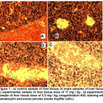 |
Figure 1(a): Control sample of liver tissue; b) sham samples of liver tissue; c) experimental sample of liver tissue dose of 17 mg / kg ; d) experimental sample of liver tissue dose of 5.5 mg / kg; (magnification 400; staining with hematoxylin and eosin) (arrows shows Kupffer cells.) Click here to View figure |
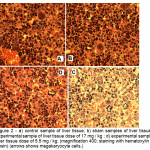 |
Figure 2(a): Control sample of liver tissue; b) sham samples of liver tissue; c) experimental sample of liver tissue dose of 17 mg / kg ; d) experimental sample of liver tissue dose of 5.5 mg / kg; (magnification 400; staining with hematoxylin and eosin) (arrows shows megakaryocyte cells.) Click here to View figure |
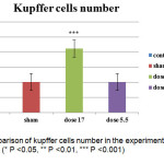 |
Figure 3: Comparison of kupffer cells number in the experimental, sham and control groups (* P <0.05, ** P <0.01, *** P <0.001) Click here to View figure |
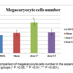 |
Figure 4: Comparison of megakaryocyte cells number in the experimental, sham and control groups (* P <0.05, ** P <0.01, *** P <0.001). Click here to View figure |
Conclusions
Nanotechnology is a technology to shrink the size of materials from 1 to 100 nm and working with materials, devices and systems on this scale. The materials at nanometer scale have very different properties from their previous properties.23 Significant increase in the use of nanomaterials has needed more attention to the benefits and risks of nanomaterials.24 wang and colleagues in a study on the effect of nano graphene oxide in mice by intravenous injection was observed that the GO did not show a clear toxicity at low doses.(GO toxicity at low doses does not show clear) and at high doses causes chronic toxicity and accumulation in the liver and lungs, and often can`t be cleared from the body by kidneys.22 The survey was conducted in 2008 by Heinlaan found that kupffer cells of liver play central role in removing nanoparticles from the bloodstream.25 Also in this paper, we study the effect of nano graphene oxide to 6 days after injection, we observed clearly increased of kupffer cells to clean liver from nano graphene oxide. Vallabani and colleagues studied the effect of carbon nanotubes on the lungs of mouse and observed at high dose mouse`s weight were significantly less than the control group, swelling and inflammation in the lungs, liver and spleen also were observed in the groups treated with higher doses.21 As the research of graphene oxide at low doses have less impact on the evolution of the liver; In another study by Huczko and colleagues in 2011 on the effect of nano graphene oxide on human lung cells observed that the concentration and exposure time are two important factors for graphene oxide nanoparticles and found that graphene oxide is toxic in high doses to lung cells and causes apoptosis in them and whatever the duration of exposure to nanoparticles increases, damaging effects caused by nanoparticles is more.26 Study of Abdel Warith and colleagues in 2011 on the liver exposed to Zn for a week, displayed various changes including degeneration of the liver cells and the natural shape of the liver was significantly disturbed and sinusoids were dilated and compressed and significantly increase of kupffer cells observed.27 In another study, Lin et al., in 2009 and Xia et al., in 2008 on the liver of mice treated with zinc oxide nanoparticles, edema and degeneration was observed in hepatocytes and Stated that the toxicity of zinc oxide nanoparticles depends on the dose and duration of exposure and its mechanism is done by Lipid peroxidation, cellular membrane damage, oxidative stress, and oxidative DNA damage.28
In this study, the number of cells involved in the inflammatory response, including megakaryocyte cells and kupffer cells increased which shows nano graphene oxide can be affected during the initial development, on the several factors during development of liver. Among these factors the number of kupffer cells and megakaryocyte cells in the liver can be noted. The impact of graphene oxide was negligible at low doses. Increasing the number of kupffer cells and megakaryocyte cells indicated toxicity and external factors related to nanomaterials in the liver. In conclusion we can say that the nano-graphene oxide can impair growth and development in the early stages of liver development in the mouse embryo.
Acknowledgment
Whereas from all members of the Department of Biology, college of sciences, Islamic Azad University, Science and Research Branch, Tehran for providing facilities executive would be appreciated.
References
- Murr L., Bang J., Guerrero P., Lopez D.,(2004), “Carbon nanotubes, nanocrystal forms and complex nanoparticle aggregates in common fuel-gas combustion source and ambient air”. J Nanoparticle Res., 5(1).
- Amato I., ( 2001),”The soot that could change the world”. FortunMagazine.
- Crispe I.N., (2009), “The liver as a lymphoid organ”. Annual Review of Annual Review of Immunology, 27, pp.147–63.
- Bilzer ,Roggel F., Gerbes A.L., (2006) “Role of Kupffer cells in host defense and liver disease”. Liver Int, 26, pp.1175–1186.
- Branehog I., Ridell B., Swolin B., Weinfeld A., (1975), “Megakaryocyte quantifications in relation to thrombokinetics in primary thrombocythaemia and allied diseases”. Scand. J. Haematol., 15 (5), pp.321–332.
- Long M.W., Williams N., and Ebbe S.,(1982),”Immature megakaryocytes in the mouse:physical characteristics, cell cycle status, and in vitro responsiveness to thrombopoietic stimulatory factor”. Blood, 59, pp.569–575.
- Morita Y., Iseki A., Okamura S., Suzuki S., Nakauchi H., and Ema H., (2011),”Functional characterization of hematopoietic stem cells in the spleen”. Hematol, 39, pp. 351–359.
- Kellie R.M. and Joseph E. I.J., (2013), “The incredible journey: From megakaryocyte development to platelet formation “. J. Cell Biol., 201(6), pp.785–796.
- Feng L.Z., Liu Z.A., (2011),”Graphene in biomedicine: Opportunities and challenges”. Nanomedicine, 6(2), pp. 317-324.
- Geim A. K., (2009),”Graphene: Status and prospects”.Science, 324(5934),pp. 1530-1534.
- Wang Y., Li Z., Wang J., Li J., Lin Y.,(2011),”Graphene and graphene oxide: Biofunctionalization and applications in biotechnology”. Trends in Biotechnology, 29(5), pp. 205-212.
- Geim A. K., Novoselov K. S.,( 2007), “The rise of graphene”.Nat. Mater., 6,pp.183- 191.
- Zhuang L., Robinson J.T., Tabakman S.M., Yang K. and Dai H., (2011). “Carbon materials for drug delivery and cancer therapy”. Materials today, 14, pp. 316-323.
- Li X., Wang X., Zhang L., Lee S., Dai H.,(2008),”Chemically derived, ultrasmooth graphene nanoribbon semiconductors”. Science, 319(5867),1229-1232.
- Dikin D. A., Stankovich S., Zimney E. J., Piner R. D., Dommett G. H. B., Evmenenko G., Nguyen S. T., Ruoff R. S.,(2007),”Preparation and Characterization of Graphene Oxide Paper”. Nature, 448, pp. 457–460.
- Stankovich S., Dikin D.A., Dommett G. H. B., Kohlhaas K. M., Zimney E.J., Stach E.A., Piner R. D., Nguyen S. T., Ruoff R. S.,(2006),” Graphene-Based Composite Materials”. Nature, 442, pp. 282–286.
- Park S. Y., Park J., Sim S. H., Sung M.G., Kim K. S., Hong B.H., (2011), “Enhanced differentiation of human neural stem cells into neurons on graphene. Advanced Materials, 23(36), 263-267.
- Stoller M. D., Park S. J., Zhu Y. W., An J.H., Ruoff R. S.,( 2008), “Graphene-Based Ultracapacitors”. Nano Lett., 8(10), pp. 3498–3502.
- Chiu W., John T., James R., Robert L., (2004),”Pulmonary Toxicity of Single-Wall Carbon Nanotubes in Mice 7 and 90 Days after Intratracheal Instillation”. Toxicological sciences, 77, pp. 126-134.
- Zhang, Yin J., Peng C., Hu W., Zhu Z.H., Li W., Fan C., Huang Q., (2011), “Distribution and biocompatibility studies of graphene oxide in mice after intravenous administration”. Carbon, 49(3), pp. 986-995.
- Vallabani N. V., Mittal S., Shukla R. K., Pandey A. K., Dhakate S. R., Pasricha R., Dhawan A., (2011), “Toxicity of Graphene in Normal Human Lung Cells (BEAS-2B)”.Biomedical Nanotechnology,7(1),pp. 106-107.
- Wang K., Ruan J., Song H., Zhang J., Wo Y., Guo S., Cui D.,(2011), ”Biocompatibility of graphene oxide”. Nanoscale Res. Lett., 6, pp.8:1–8:8.
- RafieiTabar H., (2004), “Nanotechnology and its applications in medicine and pharmacy”. Research Journal of The College of Medicine, Shahid Beheshti University of Medical Sciences and Health Services, 2, pp.111-115 [In Persian].
- Mansoori G.A., and Fauzi Soelaiman T. A., ( 2005), “ Nanotechnology–An Introduction for the Standards Community”. Journal of ASTM International, 2 (6).
- Heinlaan M., Ivask A., Blinova I., Dubourguier H.C., Kahru A.,(2008),” Toxicity of nanosized and bulk ZnO, CuO and TiO2 to bacteria Vibrio fischeri and crustaceans Daphnia magna and Thamnocephalus platyurus”. Jchemosphere, 71, pp.1308-1316.
- Huczko A., Lange H., Bystrzejewski M., Baranowski P., Jaworska H.J., Nejman P., PrzybyÅ‚owski T., Czumin´ska K., Glapin´ ski J., Walton D.R.M. and Kroto H.W., (2005), “Pulmonary Toxicity of 1-D Nanocarbon Materials”. Fullerenes, Nanotubes, and Carbon Nanostructures, 13(2), pp. 141–145.
- Abdel-Warith, A. A., Younis, E. M., Al-Asgah, N. A. and Wahbi, O. M., (2011), “Effect of zinc toxicity on liver histology of Nile tilapia,Orechromis niloticus”. Res.Essays., 6(17), pp. 3760-3769.
- Lin W., Xu Y., Huang C., Ma Y., Shannon K. B., Chen D. and Huang Y., (2009), “Toxicity of nano- and micro-sized ZnO particles in human lung epithelial cells”. Journal of Nanoparticles Research, 11, pp.25–39.